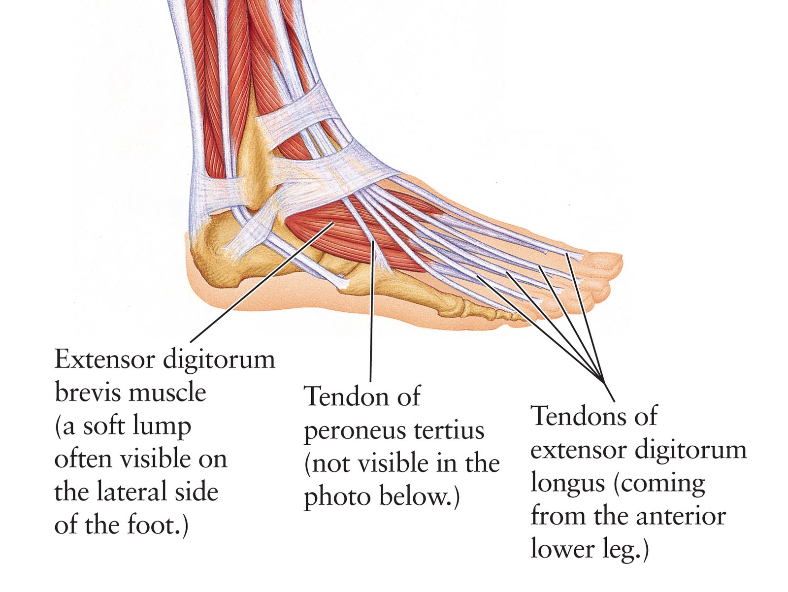
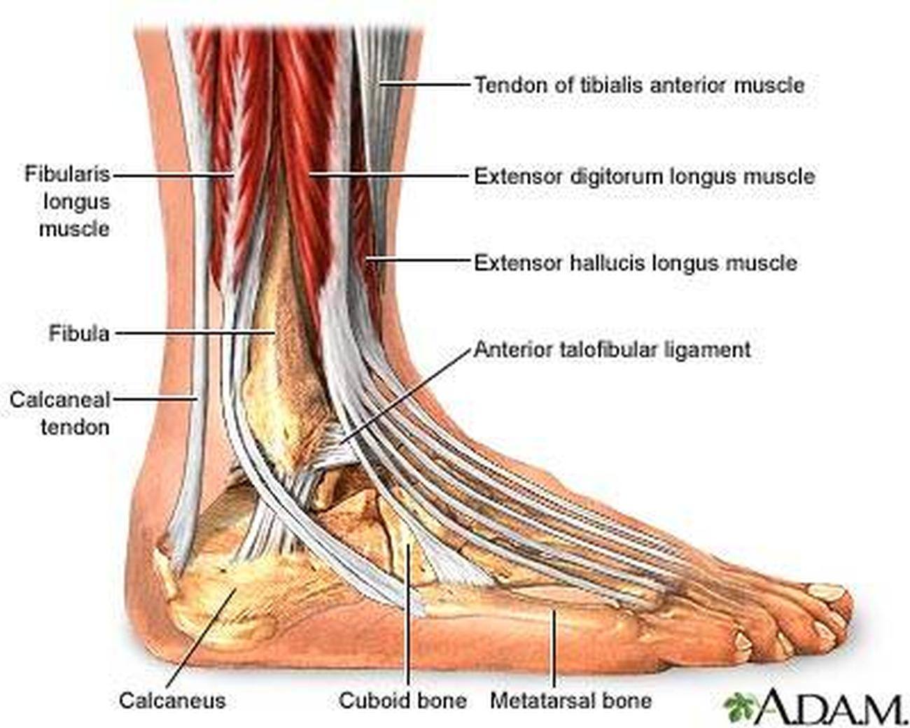
Human Anatomy for the Artist The Dorsal Foot How Do I Love Thee? Let Me Count Your Tendons
The foot is a complex mechanical structure of the human body composed of 33 joints, 26 bones, and more than a hundred muscles, tendons, and ligaments that all work together to bear weight, allow for locomotion, and transmit force. The joints of the foot are made wherever two or more of the foot bones meet.
Tendons of the Foot JOI Jacksonville Orthopaedic Institute
Tendons in the Foot Diagram: Understanding the Anatomy and Function. The foot is a complex structure composed of bones, muscles, ligaments, and tendons. While all these components work together to support our body weight and facilitate movement, it's the tendons in the foot that play a crucial role in transmitting the force generated by the.

Tendons of the Foot and Ankle TrialExhibits Inc.
Fig 1 - The dorsal layer of foot muscles. Plantar Aspect There are ten intrinsic muscles located in the plantar aspect (sole) of the foot. They act collectively to stabilise the arches of the foot and individually to control movement of the digits. They are innervated by the medial or lateral plantar nerves - which are branches of the tibial nerve.

Foot Description, Drawings, Bones, & Facts Britannica
Common causes of foot pain include plantar fasciitis, bunions, flat feet, heel spurs, mallet toe, metatarsalgia, claw toe, and Morton's neuroma. If your feet hurt, there are effective ways to ease the pain. Some conditions specific to the foot can cause pain, less movement, or instability. Verywell / Alexandra Gordon.
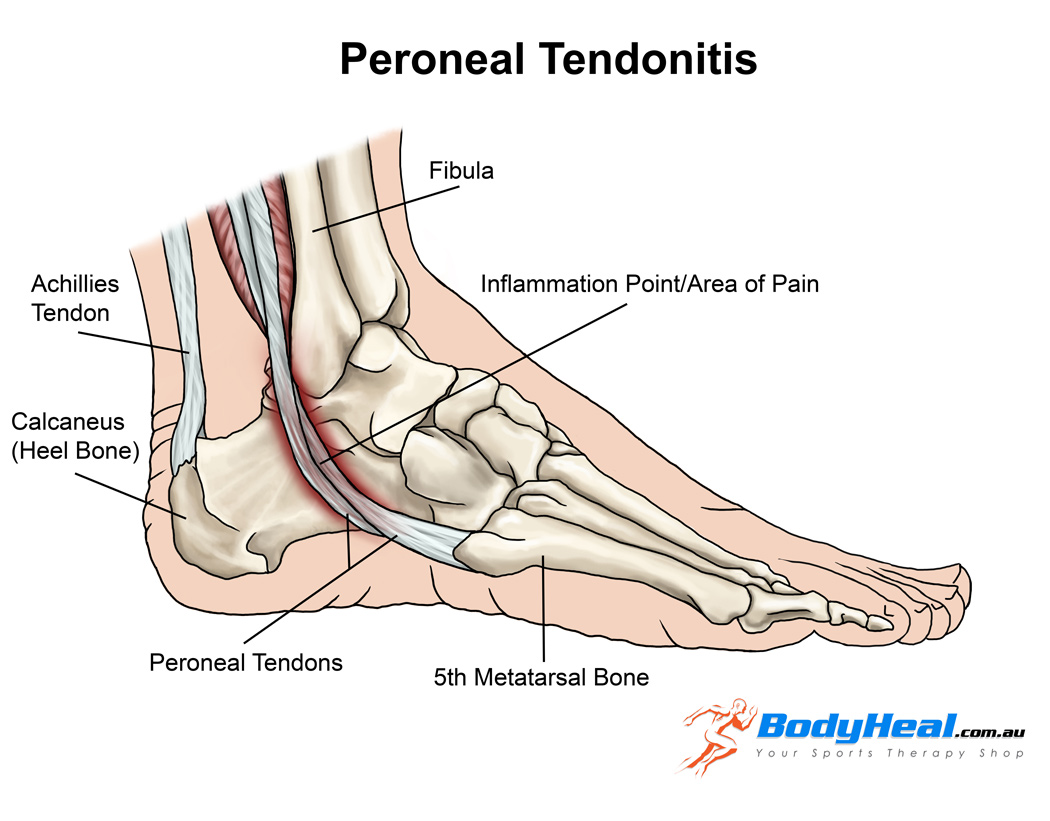
Peroneal tendinopathy, PhysioNow, Mississauga, Physiotherapy
Foot Anatomy The foot contains 26 bones, 33 joints, and over 100 tendons, muscles, and ligaments. This may sound like overkill for a flat structure that supports your weight, but you may not realize how much work your foot does!

Diagram showing the tendons and ligaments of the ankle and foot... Download Scientific Diagram
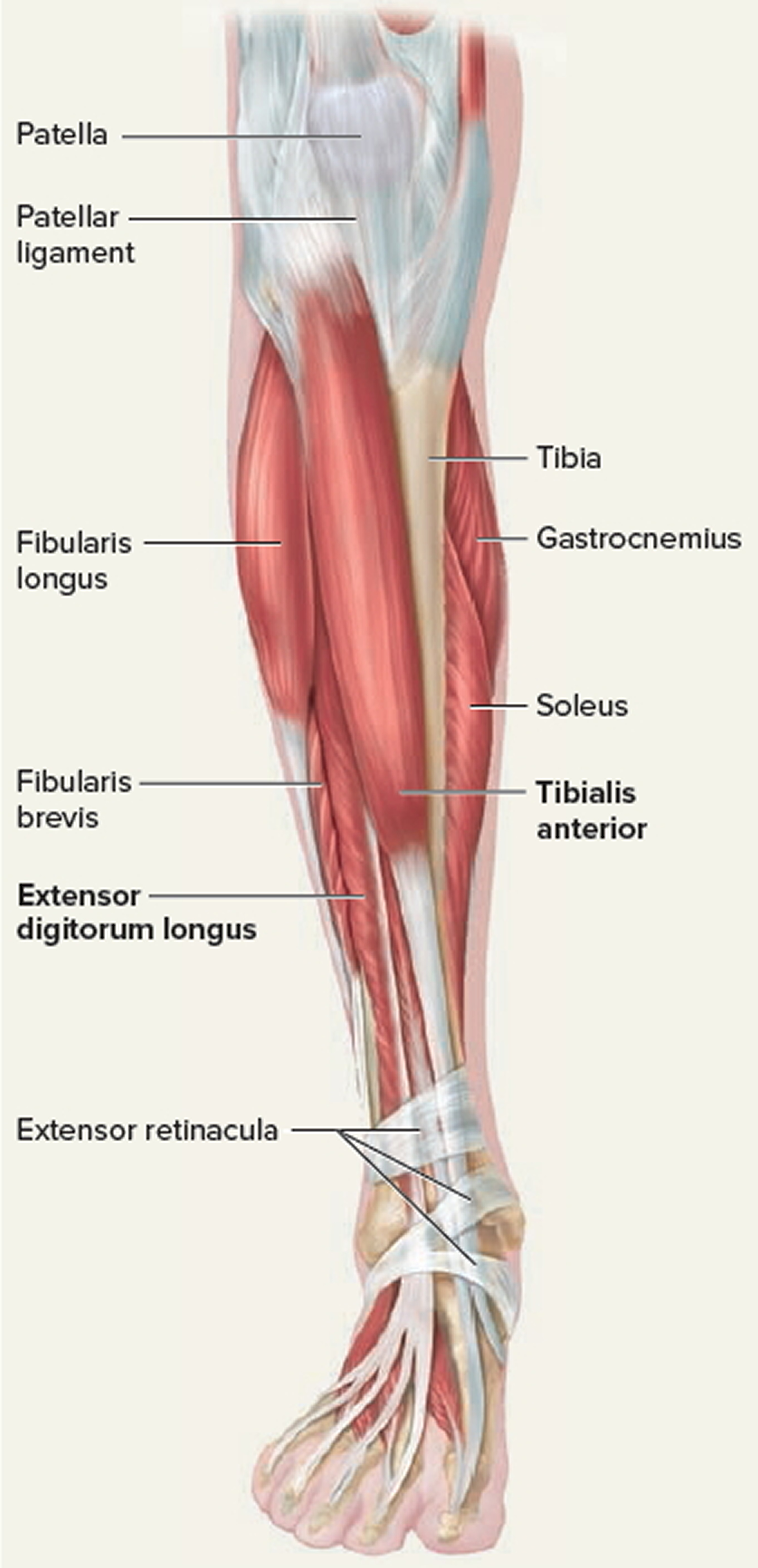
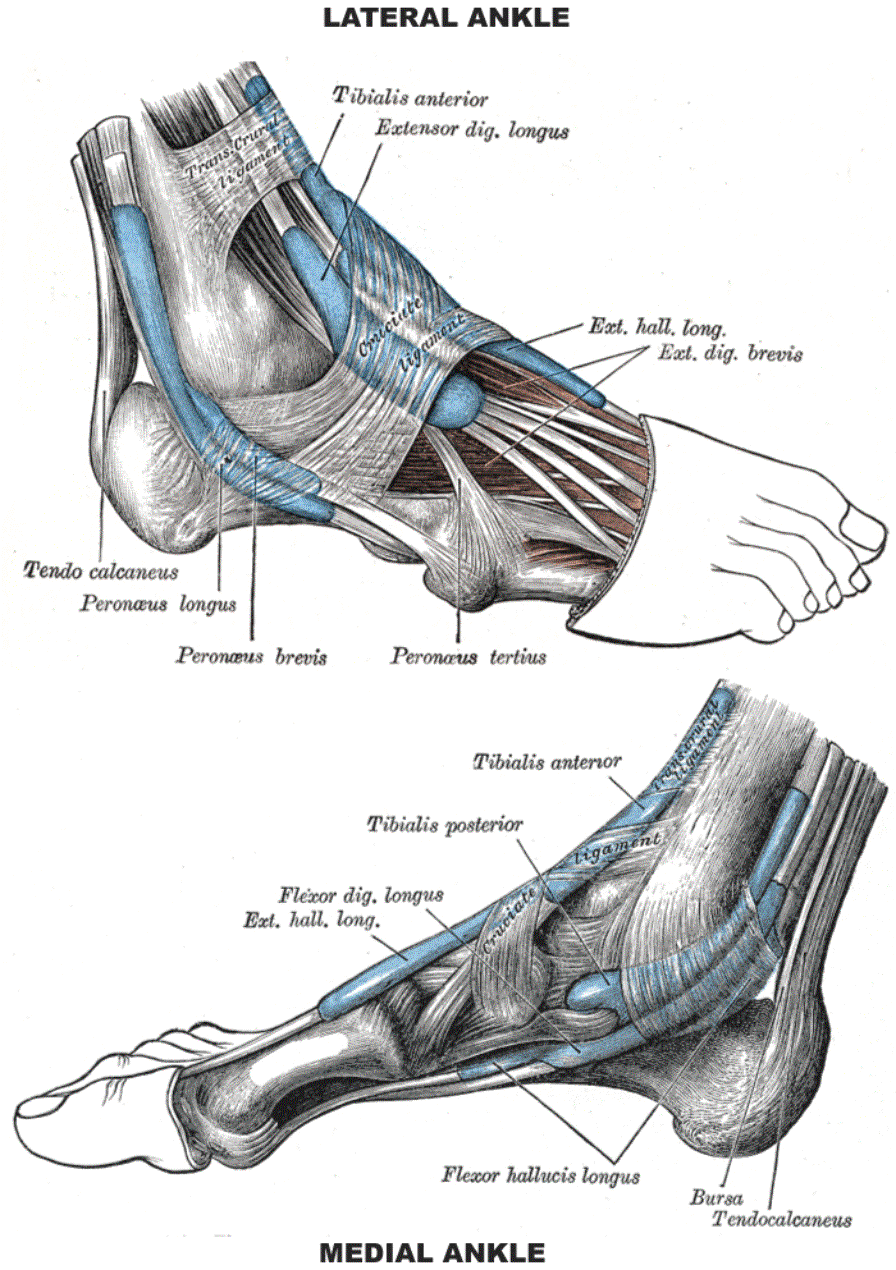
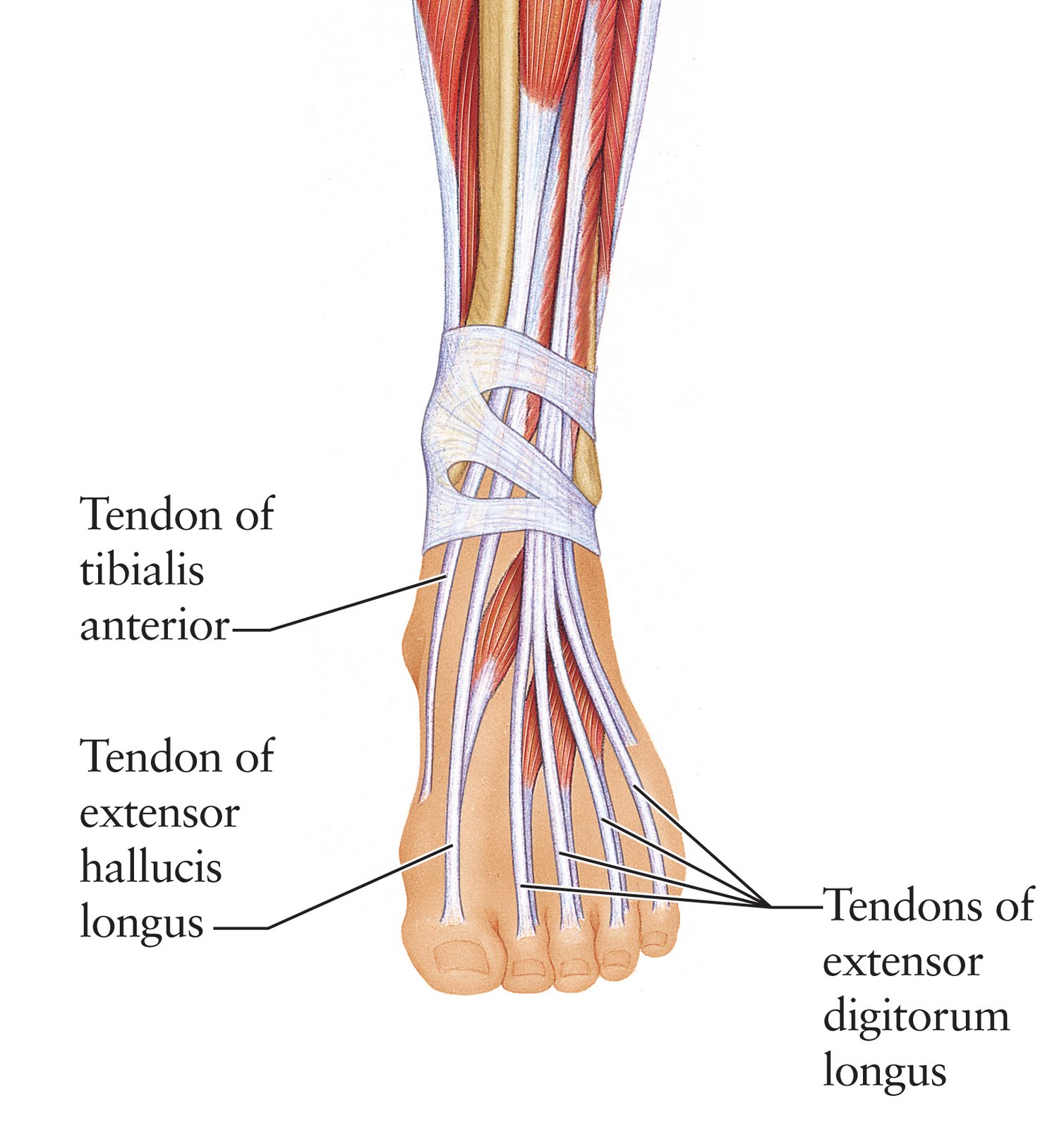
1. Tibialis Anterior Tendon The tibialis anterior muscle originates from the outer side of the tibia and passes down the front of the shin. The muscle turns into tendon about two thirds of the way down the shin and travels across the front of the ankle joint to the inner side of the foot underneath the medial foot arch.
Tendon Function, Arm, Hand Tendons Leg and Achilles Tendons
Structure of the foot kool99/Getty Images In the foot, there are: 26 bones 33 joints more than 100 muscles, tendons, and ligaments Bones of the foot The bones in the foot make up nearly.
Pictures Of Ankle Joint Ligaments
Foot and ankle anatomy consists of 33 bones, 26 joints and over a hundred muscles, ligaments and tendons. This complex network of structures fit and work together to bear weight, allow movement and provide a stable base for us to stand and move on.
Foot (Anatomy) Bones, Ligaments, Muscles, Tendons, Arches and Skin
Anatomy is a road map. Most structures in the foot are fairly superficial and can be easily palpated. Anatomical structures (tendons, bones, joints, etc) tend to hurt exactly where they are injured or inflamed.
Extensor Tendons Of Foot Photograph by Microscape/science Photo Library Fine Art America
The anatomy of the foot The foot contains a lot of moving parts - 26 bones, 33 joints and over 100 ligaments. The foot is divided into three sections - the forefoot, the midfoot and the hindfoot. The forefoot This consists of five long bones (metatarsal bones) and five shorter bones that form the base of the toes (phalanges).
Strained Peroneal Tendon...? run.around.aroo
Products Human body Muscular System Muscles Muscles The 20-plus muscles in the foot help enable movement, while also giving the foot its shape. Like the fingers, the toes have flexor and.
Ankle Ligaments And Tendons Anatomy Picture Reference and Health News
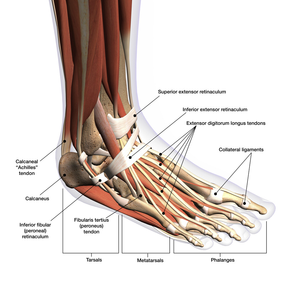
Ankle joint Explore study unit Joints and ligaments of the foot Explore study unit Bones of the foot There are 26 bones in the foot, divided into three groups: Seven tarsal bones Five metatarsal bones Fourteen phalanges Tarsals make up a strong weight bearing platform.
anatomy of the foot tendons
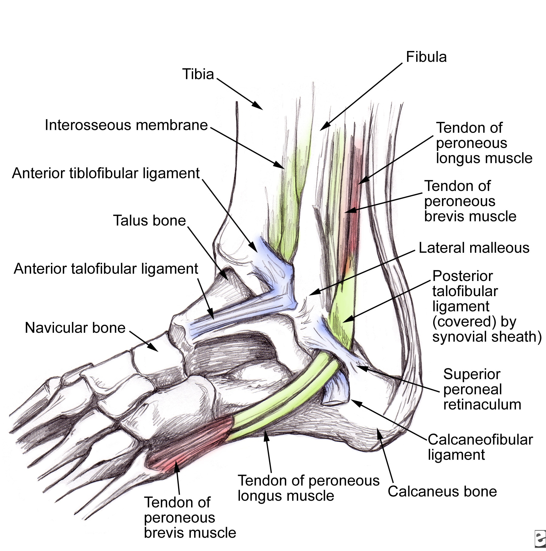
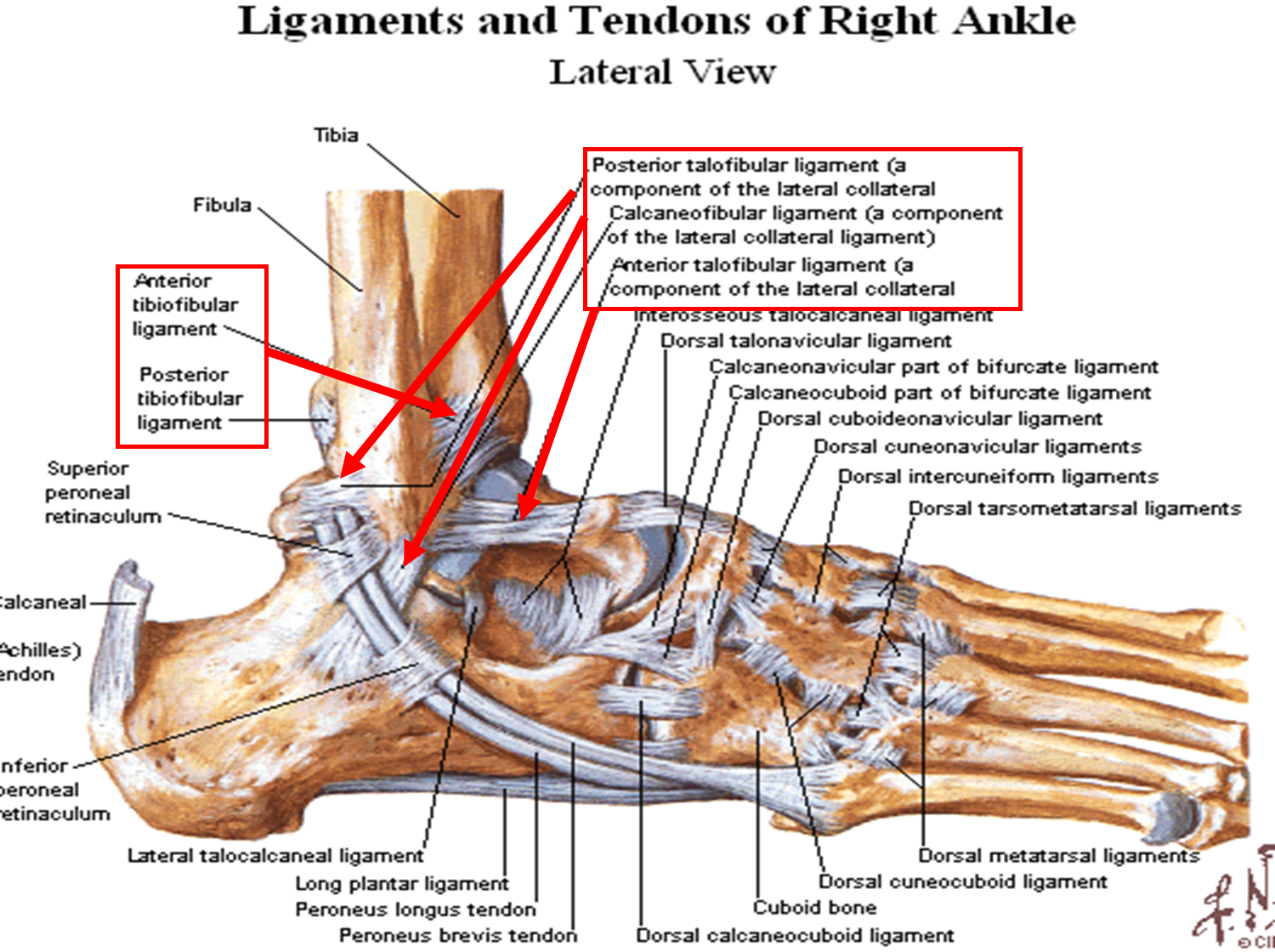
The ankle consists of three bones attached by muscles, tendons, and ligaments that connect the foot to the leg. In the lower leg are two bones called the tibia (shin bone) and the fibula. These bones articulate (connect) to the Talus or ankle bone at the tibiotalar joint (ankle joint) allowing the foot to move up and down.

Peroneal Tendons What are they? Best treatment options in 2023
Overview What is foot tendonitis? Foot tendonitis (tendinitis) is inflammation or irritation of a tendon in your foot. Tendons are strong bands of tissue that connect muscles to bones. Overuse usually causes foot tendonitis, but it can also be the result of an injury. Are there different types of foot tendonitis? Your feet contain many tendons.
Human Anatomy for the Artist The Dorsal Foot How Do I Love Thee? Let Me Count Your Tendons
It consists of 28 bones, which can be divided functionally into three groups, referred to as the tarsus, metatarsus and phalanges. The foot is not only complicated in terms of the number and structure of bones, but also in terms of its joints.

Peroneal Tendonitis DOC
What are ligaments? Ligaments are bands of fibers interconnected in strong, cord-like ropes. In your feet, ligaments attach bones to each other. You have ligaments all over your body that hold bones together. Some ligaments also support internal organs. Function What is the purpose of the foot ligaments?
