
Inferior view of the brain showing the five main visual pathways, in
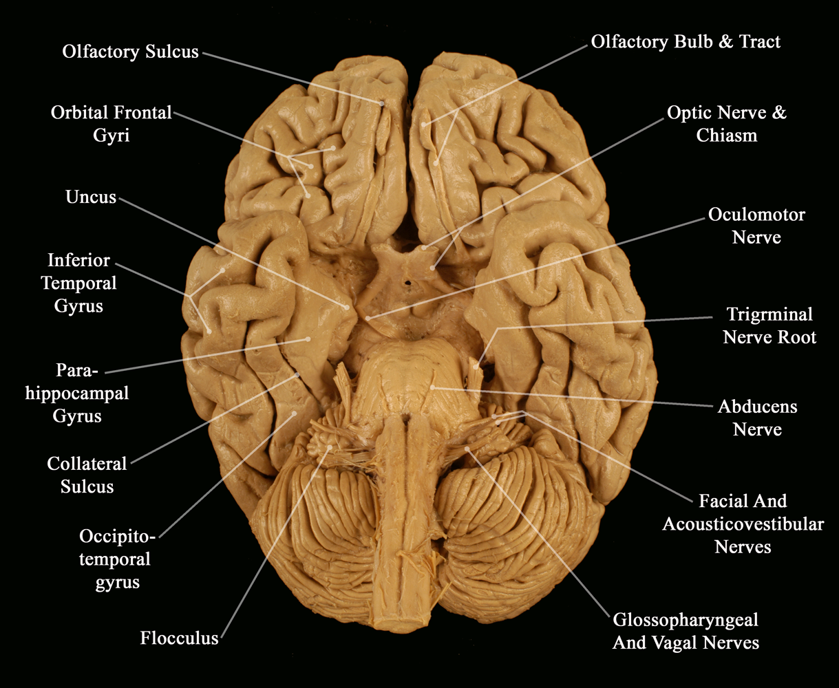
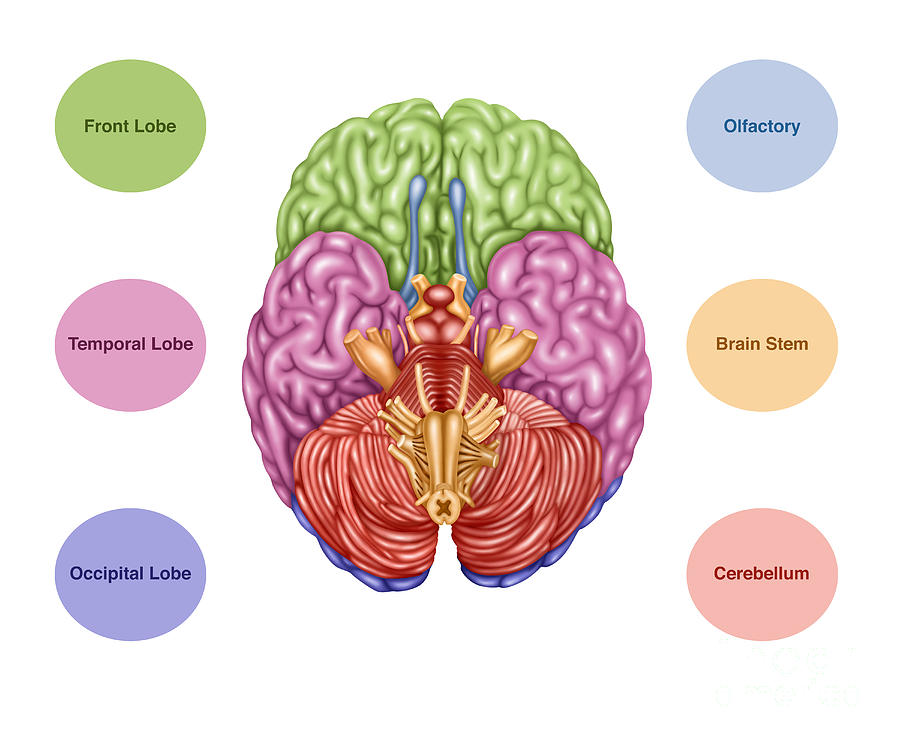
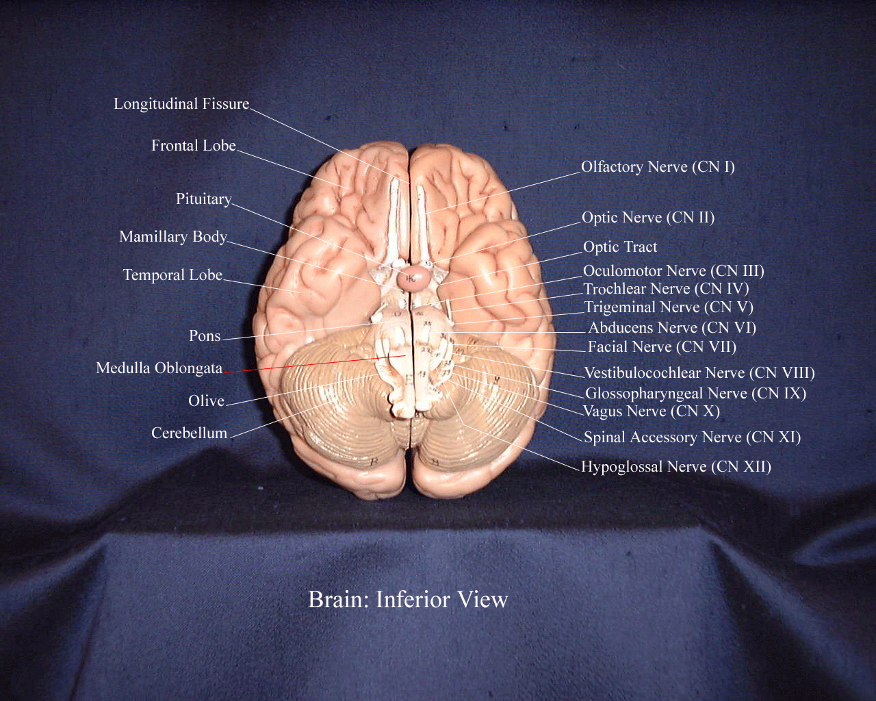
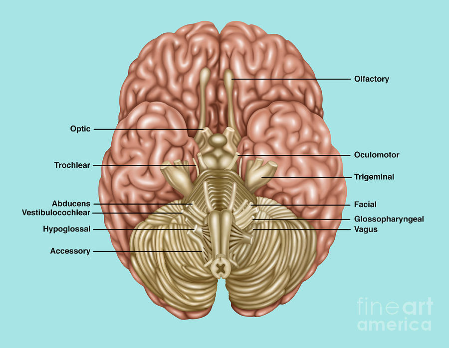
Description. This panel depicts the cranial nerves of the brain from an inferior (bottom) view. The illustration includes the following details: frontal lobe, temporal lobe, occipital lobe, cerebellum and brainstem, olfactory nerve (I), optic nerve (II), oculomotor nerve (III), trochlear nerve (IV), trigeminal nerve (V), abducens nerve (VI), facial nerve (VII), vestibulocochlear nerve (VIII.
Cerebrum Overview
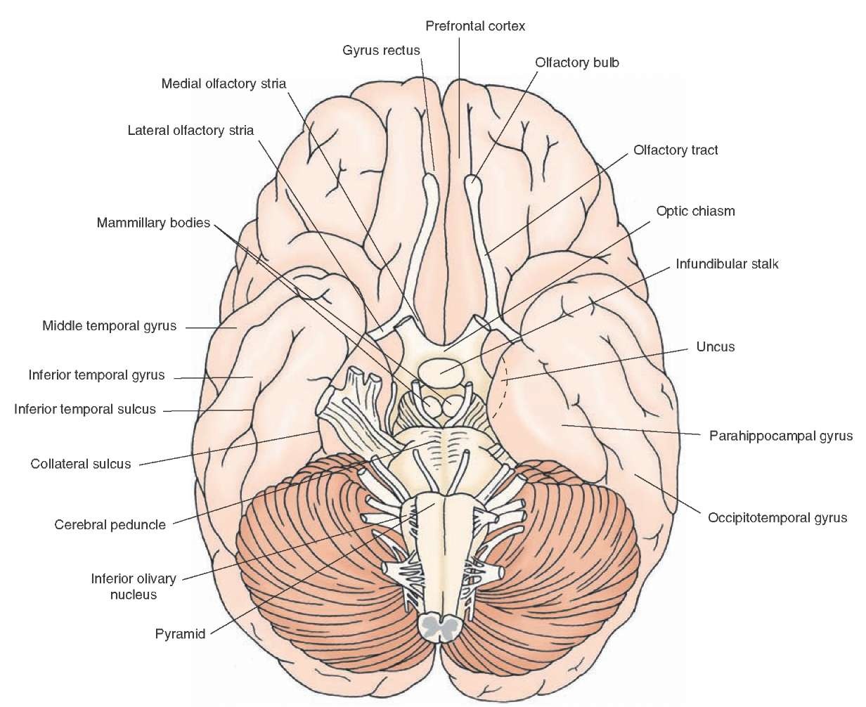
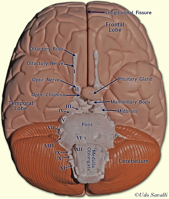
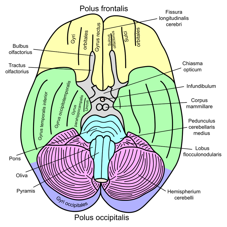
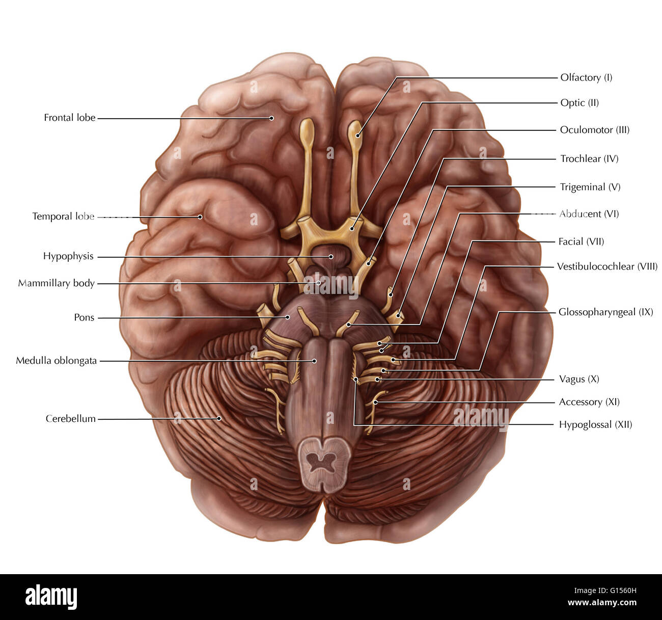
Above: Lateral view of the brain stem showing the locations of the cranial nerves III - XII. The, olfactory nerves (I) and optic nerves (II) emerge from the cerebrum or forebrain, and the remaining ten pairs arise from the brainstem, which is the lower part of the brain. Above: Inferior view of the brain with the pairs of cranial nerves labeled.

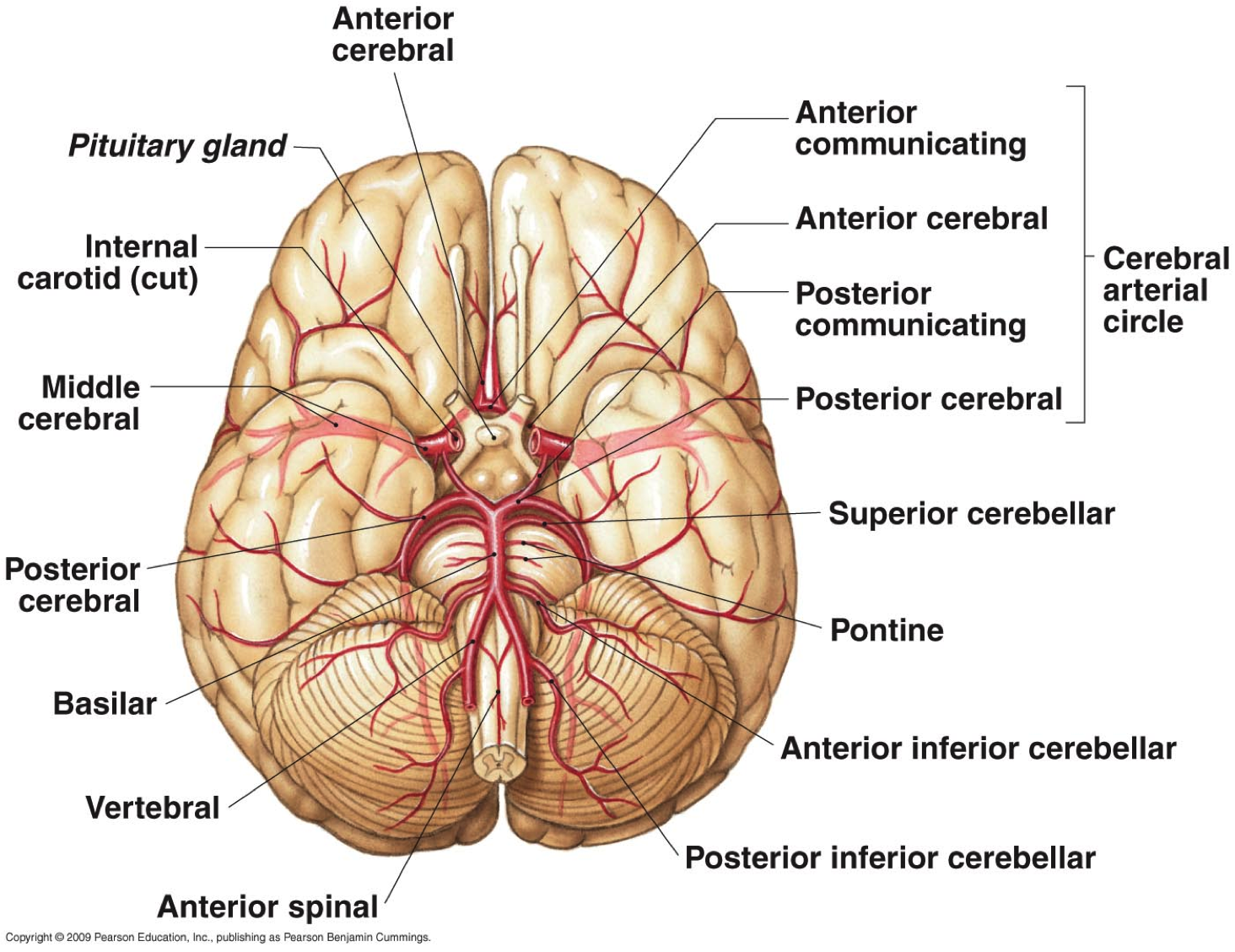
7 Inferior view of arteries of the brain. Circle of Willis is depicted
ID: 71551 Title: Inferior View of the Cere… Category: Labeled ID: 68294 Title: Inferior Surface of Brain Category: Labeled -

Brain 🧠 inferior view Medical school studying, Nursing school
Anatomy of the Brain There are different ways of dividing the brain anatomically into regions. Let's use a common method and divide the brain into three main regions based on embryonic development: the forebrain, midbrain and hindbrain. Under these divisions:
Overview of the Central Nervous System (Gross Anatomy of the Brain) Part 2
The sagittal view of the midbrain reveals its two portions: tectum and tegmentum. The tectum is the region of the midbrain posterior to the cerebral aqueduct of Sylvius. It is composed of the two posterior bulges called the superior and inferior colliculi. They are involved in processing of visual and auditory stimuli respectively.
BIO201Human Brain
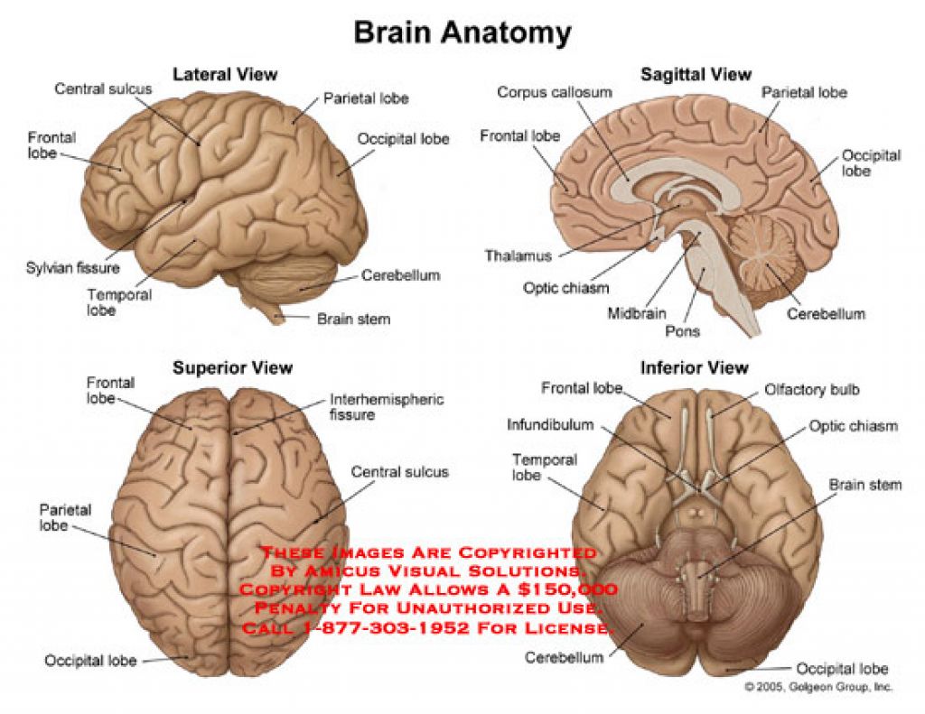
1 2 Each point of view provides an altered perspective of the brain that changes the appearance of the major divisions, landmarks, and structures. Anatomical directions 1 2 3 4 Next Quickly learn the parts of the brain with these interactive quizzes and labelling exercises. Reference planes: 1 2 3 Views of the brain: 1 2 3 4 5 6
Introduction to Neuroanatomy Physiopedia
Inferior view of the brain 5.0 (1 review) Get a hint Frontal lobe Click the card to flip 👆 What is 1 Click the card to flip 👆 1 / 12 Flashcards Learn Test Match Q-Chat Created by lindamed331 Students also viewed Chapter 11 49 terms calia_meads Preview Eye & Ear Diagram Labeling 22 terms Sydney_Walker398 Preview Ch 12 HW - Mastering A&P 45 terms
Brain Anatomy, Inferior View Photograph by Gwen Shockey Fine Art America
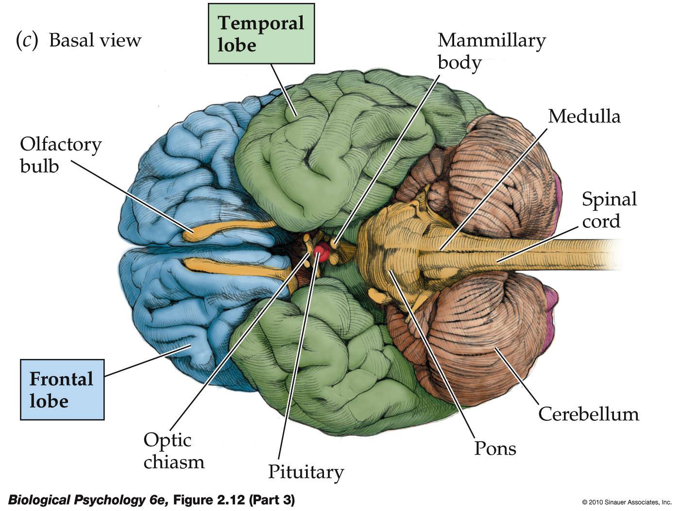
The inferior surface of the base of the brain (basis), with its arteries. Translated by: Ronald A. Bergman, PhD and Adel K. Afifi, MD, MS Peer Review Status: Internally Peer Reviewed Magnified View (via Quicktime VR) A. Anterior cerebral frontal lobe [OBS]. B. Middle cerebral temporal lobe [OBS]. C. Posterior occipital lobe [OBS]. D. Cerebellum. E.
Brain Diagram Cliparts.co
The brain (Latin: cerebrum) is the central anatomical part of the nervous system, and it is located in the cranial cavity of the skull. The brain is made up of the cerebrum, diencephalon, brainstem and cerebellum. It is a complex organ composed of neural tissue.
Life After Being A Student My Mission To Learn The Brain
Diencephalon Inferior view Frontal lobe Temporal lobe Highlights Lateral view Medial view Inferior view Sources + Show all

inferior view of brain Diagram Quizlet
Brain inferior view Stock Photos and Images (234) See brain inferior view stock video clips Quick filters: Cut Outs | Vectors | Black & white Sort by Relevant RF 2BEH8CG - Brain Anatomy, Inferior View, Illustration RF FPR641 - Anatomy of human brain, inferior view.
InferiorBrainModel
This article will describe the anatomy from the inferior view of the skull base. We will explore the many foramina and projections that enable arteries and nerves to both enter and leave the skull.
The Neurocritic The Purring Center in Cats
1/7 Synonyms: Forebrain, Endbrain , show more. The brain, along with the spinal cord, is the main organ of the central nervous system. It is the most complex organ of the body, with many layers and components that play their roles in almost every function performed by the body. The brain is composed of the cerebrum, cerebellum and brainstem.

Brain Anatomy, Illustration Brain anatomy, Human brain anatomy, Brain
Start studying Inferior View of the Brain. Learn vocabulary, terms, and more with flashcards, games, and other study tools.. practical 1 mc questions anatomy 2 lab. 50 terms. chrisdoughh. Preview. CS-19 BLOOD : DIAGRAMS. 33 terms. ami_piddington. Preview. ch 15 hematologic system paper. 105 terms. vickygrussell.
Brain Anatomy, Inferior View Photograph by Gwen Shockey
The lateral view of the brain shows the three major parts of the brain: cerebrum, cerebellum and brainstem . A lateral view of the cerebrum is the best perspective to appreciate the lobes of the hemispheres. Each hemisphere is conventionally divided into six lobes, but only four of them are visible from this lateral perspective.
Brain Inferior View Stock Photos & Brain Inferior View Stock Images Alamy
Learn about the features, markings, and distinguishing characteristics of the brain; then test yourself with labeled images, hints, and answer keys that put you in control. Structure-Function.org. Resources biology human anatomy ☰ Brain Model, inferior view « Inferolateral view | Brain main.
