
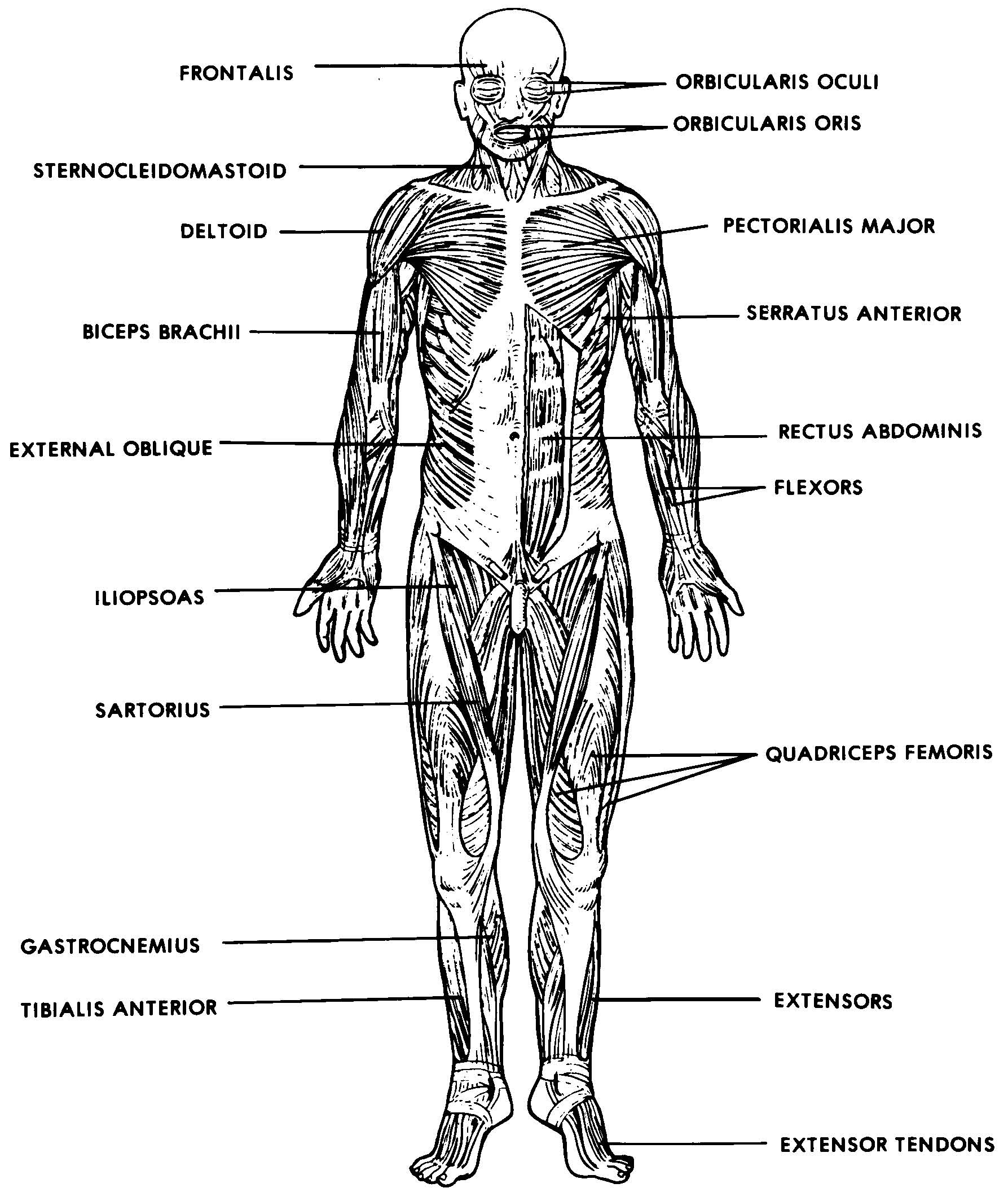
Muscles Diagrams Diagram of muscles and anatomy charts
Muscles attach to bones directly or through tendons or aponeuroses. Skeletal muscles maintain posture, stabilize bones and joints, control internal movement, and generate heat. Skeletal muscle fibers are long, multinucleated cells. The membrane of the cell is the sarcolemma; the cytoplasm of the cell is the sarcoplasm.
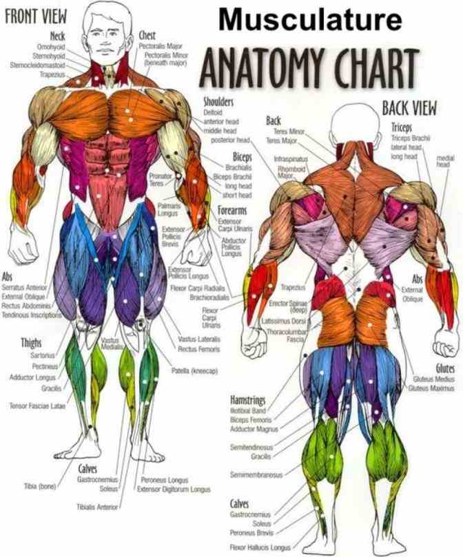
Simple Human Muscles Diagram / Learn All Muscles With Quizzes And
Gastrocnemius (calf muscle): One of the large muscles of the leg, it connects to the heel. It flexes and extends the foot, ankle, and knee. Soleus: This muscle extends from the back of the knee to.

Labeled Muscle Diagram Chart Free Download
Labelled Diagram of Skeletal Muscle . Skeletal Muscle - Description . The muscle tissue is composed of a large number of myocytes or muscle cells. These muscle cells are slender and long and are termed as muscle fibres. The skeletal muscle fibres are multinucleated. They are arranged parallel to each other, along with some intervening.

Simple Human Muscles Diagram / Learn All Muscles With Quizzes And
Leg muscles labeled. Take a look at the leg muscles diagram below, where you see each muscle clearly labeled. Spend some time revising this diagram by connecting the name and location of each structure with what you've just learned in the video. The aim of this exercise is to improve your confidence in identifying different structures.
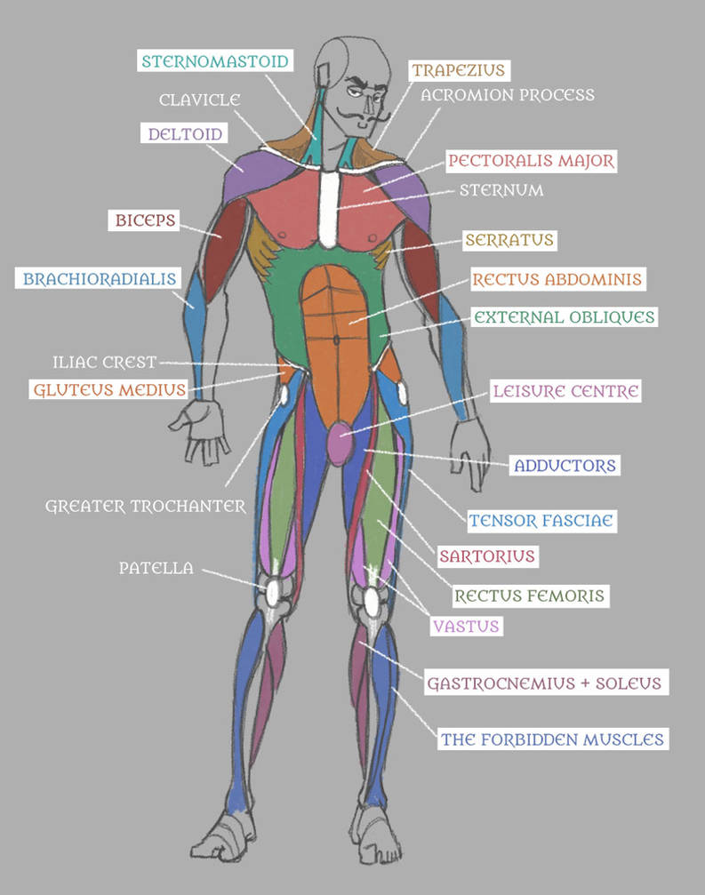
Labeled Body Muscle Diagram
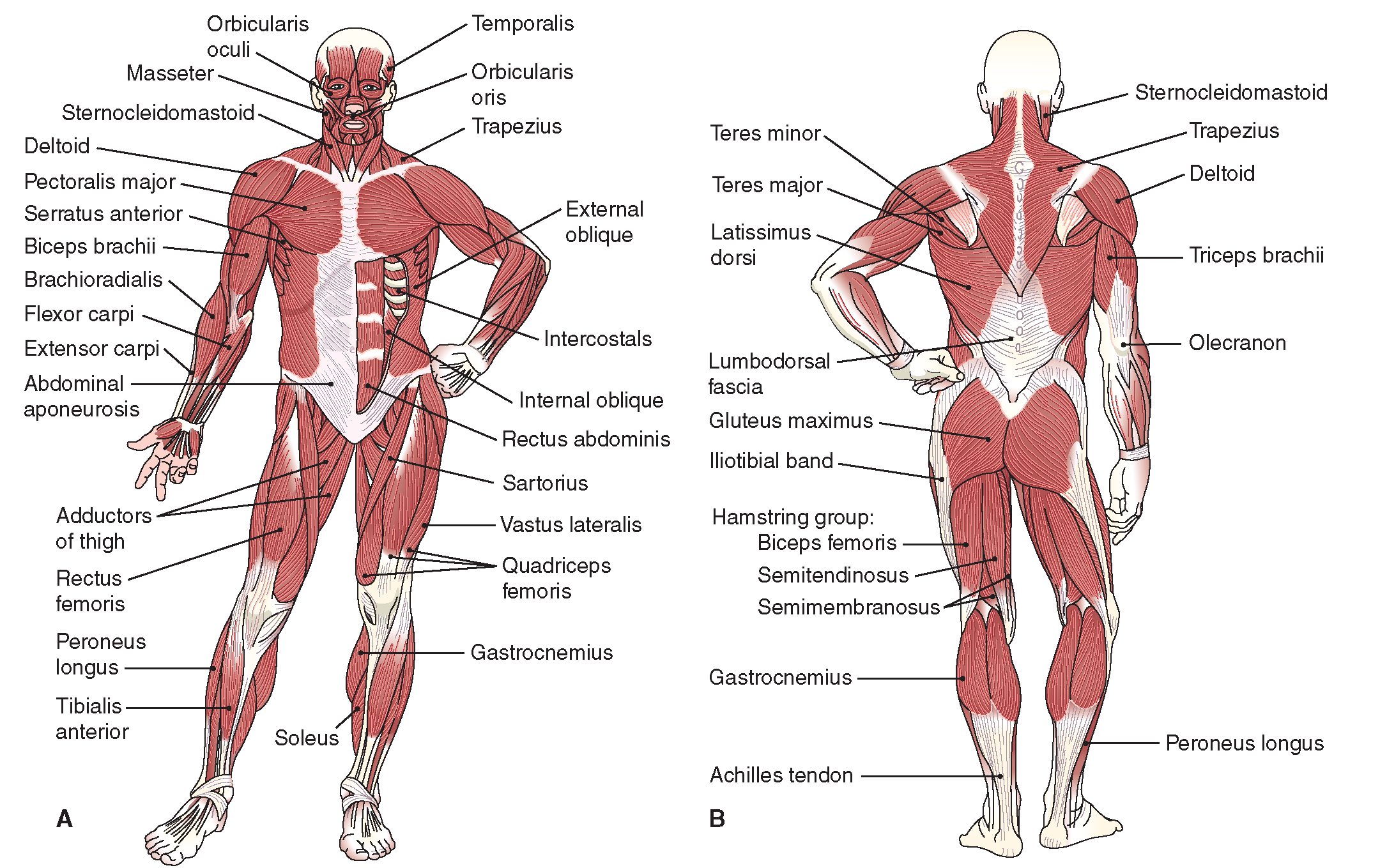
Human body muscle diagrams. Muscle diagrams are a great way to get an overview of all of the muscles within a body region. Studying these is an ideal first step before moving onto the more advanced practices of muscle labeling and quizzes. If you're looking for a speedy way to learn muscle anatomy, look no further than our anatomy crash courses .
The Musculoskeletal System (Structure and Function) (Nursing) Part 4
This nursing test bank includes questions about Anatomy and Physiology and its related concepts such as: structure and functions of the human body, nursing care management of patients with conditions related to the different body systems. Dive into the ultimate study guide for the muscular system, where anatomy and physiology converge.

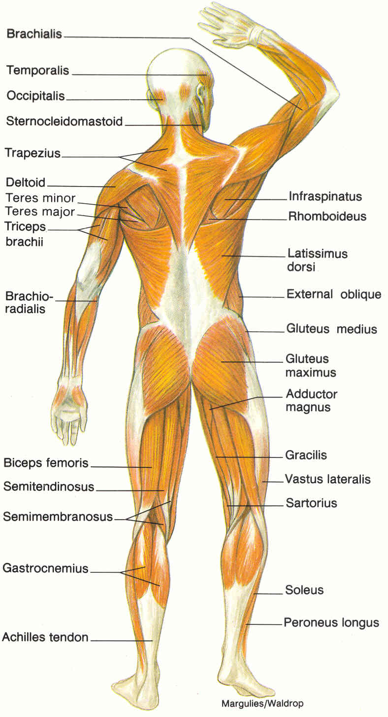
Back Muscles Anatomy Labeled Upper Back Anatomy and physiology
Attached to the bones of the skeletal system are about 700 named muscles that make up roughly half of a person's body weight. Each of these muscles is a discrete organ constructed of skeletal muscle tissue, blood vessels, tendons, and nerves. Muscle tissue is also found inside of the heart, digestive organs, and blood vessels.
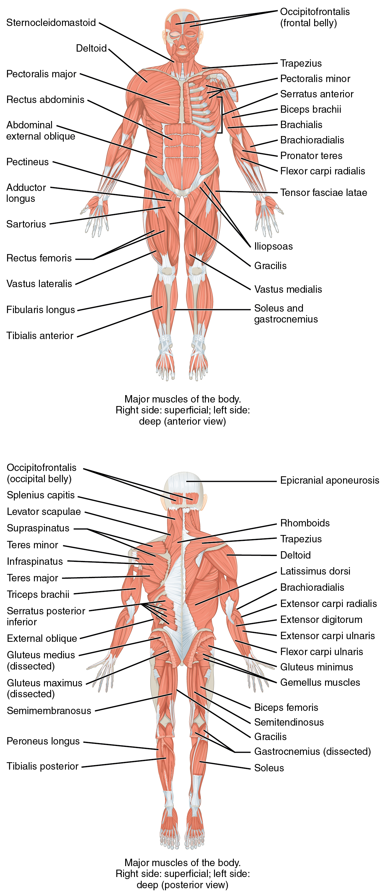
11.2 Naming Skeletal Muscles Anatomy and Physiology
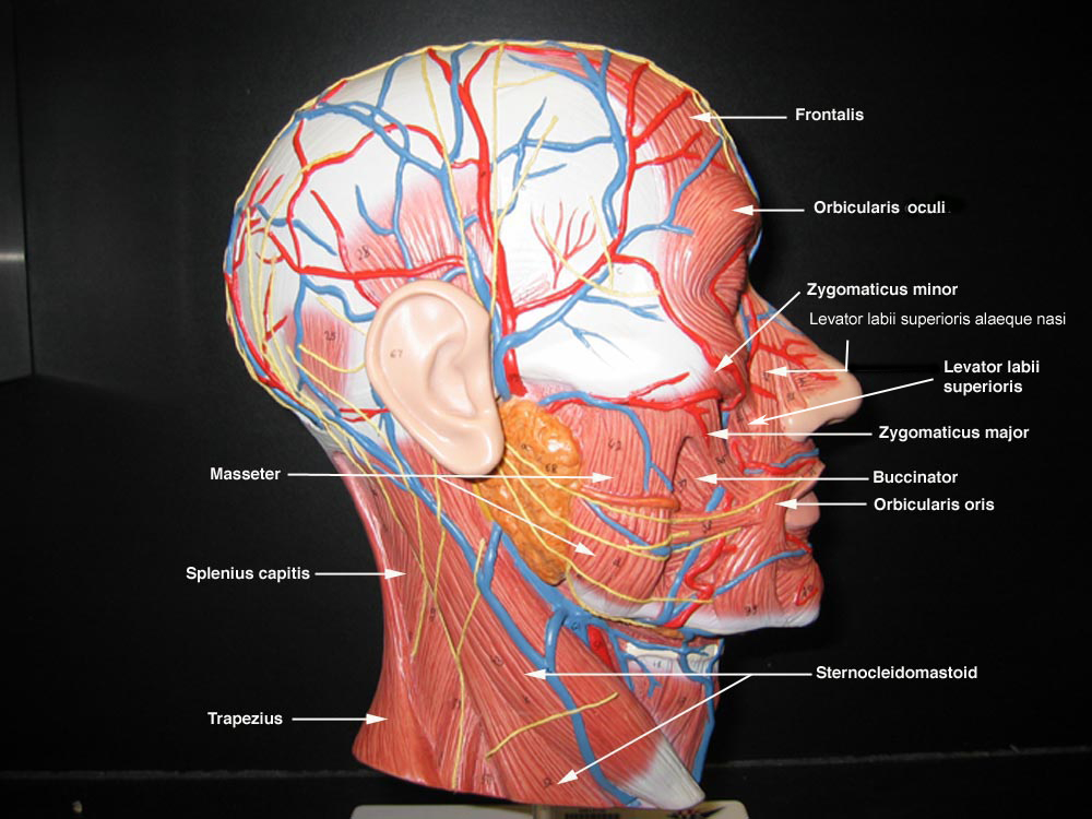
Practice test. To begin, spend a few minutes analysing the face muscles diagram above in which all the face muscles are clearly labeled. Once you think you've got a solid idea of the location of each muscle (top tip: try to associate the location with the function to aid your memory!), you can try labelling the muscles yourself using our.
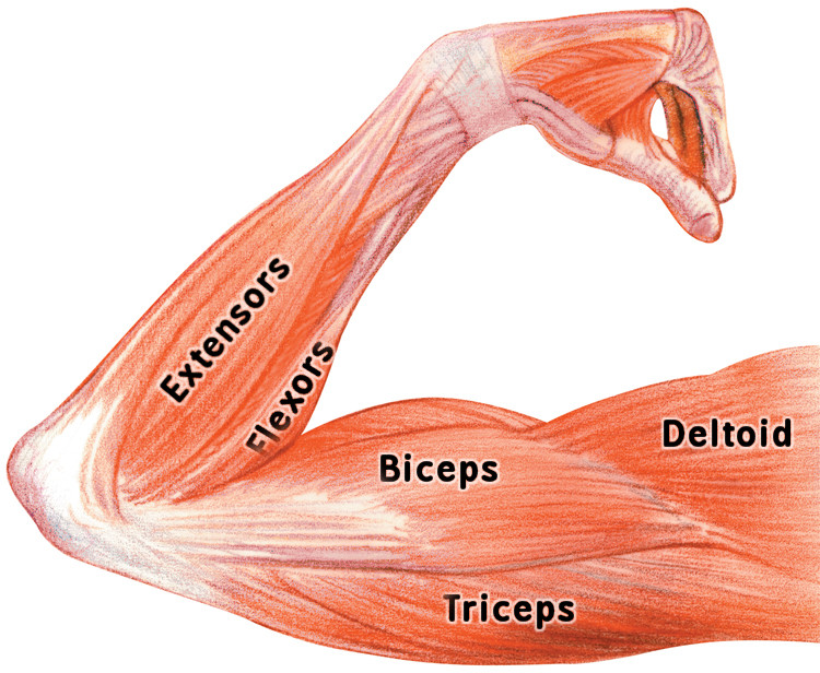
Arm Muscles Diagram Labelled Diagram Of The Muscles In The Human Body
Muscular. The primary job of muscles is to move the bones of the skeleton, but muscles also enable the heart to beat and constitute the walls of other vital hollow organs. Skeletal muscle: This.

Back Muscles Diagram Labeled Labeled Muscle Diagram — UNTPIKAPPS We
Leg, Hip & Gluteal Anatomy. Gluteal Muscles. Hamstring Muscles. Hip Adductors. Hip Flexors (Iliopsoas) Quadriceps Muscles. Neck Anatomy. Triceps Anatomy. Shoulder Anatomy (Deltoids & Rotator Cuff)
Human Anatomy Muscles with Labels! by Pseudolonewolf on DeviantArt
Striated muscle makes up the muscles in the body that are attached to the skeleton. Striated muscle is made up of muscle fibres. A muscle fibre is a highly specialised cell-like unit: Each muscle fibre contains an organised arrangement of contractile proteins in the cytoplasm. Each muscle fibre is surrounded by a cell surface membrane.

Human Body Muscle Diagrams 101 Diagrams
Each skeletal muscle is an organ that consists of various integrated tissues. These tissues include the skeletal muscle fibers, blood vessels, nerve fibers, and connective tissue. Each skeletal muscle has three layers of connective tissue (called "mysia") that enclose it and provide structure to the muscle as a whole, and also.

The Basic Muscles In The Human Body These Bones Of Mine
Inner hip & gluteal muscles. Anterior, medical and posterior thigh muscles. Anterior, lateral and posterior leg muscles. Dorsal and plantar foot muscles. This eBook contains high-quality illustrations and validated information about each muscle. It is available for free. Download free PDF (8.5MB) Get for free on iBooks.

Pin on Anatomy Diagrams
Osmosis High-Yield Notes. Muscular system anatomy and physiology. Sliding filament model of muscle contraction. Muscle contraction. Neuromuscular junction and motor unit. Osmosis Muscles high-yield notes offers clear overviews with striking illustrations, tables, and diagrams. Make learning more manageable.
Neck Muscle Diagram Labeled muscles to label Google Search Human
An extensor muscle that straightens or lifts the foot. A muscle of the leg originating on the lateral condyle of the tibia and the interosseus membrane between the tibia and the fibula and inserted in the first cunieform and first metatarsal bones. It dorsi flexes and inverts the foot, supports the arch, and is controlled by the deep peroneal.
Labelled Diagram Of Muscles In Body / Pin by ashlee brown on nursing
Muscular system functions include mobility, stability, posture, circulation, and more. Muscles allow a person to move, speak, and chew. They control heartbeat, breathing, and digestion. Other.
